by Robert Haverly | Jul 19, 2026 | News
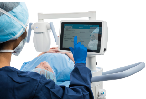
I am pleased to announce that we now offer Epioxa™ HD / Epioxa™, the first and only FDA-approved epithelium-on (epi-on) corneal cross-linking treatment for keratoconus (KC). As a Principal Investigator in the clinical trial evaluating Epioxa™, I had the opportunity to gain firsthand experience with this innovative treatment and am pleased to now offer it to patients in our region. By preserving the corneal epithelium, this oxygen-enhanced treatment promotes faster healing and significantly less discomfort than traditional epithelium-off procedures, making treatment more comfortable and accessible for many patients. I welcome you to observe a treatment in our office or attend one of our upcoming educational sessions to see the procedure firsthand.
Methodology
Epioxa is indicated for patients 13 years of age and older and is performed using the O2n™ System and Boost Goggles®. Because the procedure is incision-free and leaves the epithelium intact, it reduces many of the risks associated with epithelial removal. In addition, treatment does not require documented disease progression before intervention, allowing earlier treatment to help preserve vision and potentially reduce the need for corneal transplantation. In clinical trials, both eyes could be treated as little as one week apart.
We are now scheduling evaluations. Patients referred for Epioxa will also receive support through a dedicated Patient Access Liaison and EpioxaCareConnect™.
Who Can Benefit From Cross-linking?
Please consider referring patients with suspected keratoconus, irregular astigmatism, unexplained best-corrected vision worse than 20/20, or rapidly changing refractive error for topographic and tomographic evaluation. Earlier diagnosis allows for earlier treatment, helping preserve vision before significant progression occurs.
https://www.erielasereye.com/additional-service/keratoconus-corneal-cross-linking/
https://www.glaukos.com/int/cornea/cross-linking/

by Robert Haverly | May 25, 2026 | News
Fireworks and Eye Safety: Protecting Your Vision This Summer
Fireworks are a cherished part of summer celebrations, but every year they cause thousands of serious injuries — many involving the eyes. Despite being sold legally and often marketed as family entertainment, fireworks are explosives capable of causing permanent vision loss, severe burns, and even death.
According to the U.S. Consumer Product Safety Commission (CPSC), eight people died and nearly 14,700 people were seriously injured by fireworks in the United States in 2024 alone — a dramatic 58% increase compared with the previous year. Nearly one in five fireworks injuries involved the eyes.
Even “Safe” Fireworks Can Be Dangerous
Many people believe that legal fireworks are safe if used carefully. Unfortunately, that is not always true.
Fireworks can malfunction unexpectedly, discharge in the wrong direction, or explode all at once. Even experienced, safety-conscious users can be seriously injured. Simple mistakes — such as holding a firework at the wrong angle or approaching a malfunctioning device — can have devastating consequences.
In some cases, injuries occur because the fireworks themselves are defective. The CPSC has reported that many consumer fireworks contain dangerous modifications, unsafe explosive materials, or altered fuses that increase the risk of malfunction.
The reality is that most fireworks injuries occur with legally purchased consumer fireworks.
Children and Bystanders Are Frequently Injured
One of the most concerning aspects of fireworks injuries is how often innocent bystanders are harmed. Children, family members, and spectators may suffer injuries even when they are not handling fireworks themselves.
Children age 15 and under account for nearly one-third of fireworks injuries, while teenagers ages 15 to 19 have some of the highest rates of emergency room visits related to fireworks.
Sparklers, commonly considered harmless, are especially dangerous. They burn at temperatures exceeding 2,000 degrees Fahrenheit — hot enough to melt metal. Sparklers alone are responsible for hundreds of injuries every year and can cause severe burns and blinding eye injuries within seconds.
Fireworks Can Cause Permanent Eye Damage
Fireworks-related eye injuries can be devastating. The force of an explosion combined with heat and chemicals can cause multiple forms of trauma at the same time.
Serious eye injuries may include:
- Corneal abrasions
- Chemical burns
- Thermal burns
- Retinal detachment
- Internal bleeding
- Rupture of the eyeball (globe rupture)
- Permanent vision loss or blindness
Some patients require emergency surgery, and in severe cases, vision cannot be restored.
What To Do If a Fireworks Eye Injury Occurs
An eye injury caused by fireworks is a true medical emergency. Immediate treatment may help preserve vision.
If an injury occurs:
- Do not rub the eye
- Do not rinse the eye with water
- Do not apply pressure
- Do not attempt to remove debris or objects from the eye
- Avoid aspirin or ibuprofen unless instructed by a physician
- Seek emergency medical care immediately or call 911
Fireworks Safety Tips
The safest way to enjoy fireworks is to attend a professional public display rather than using fireworks at home.
If you are around fireworks, follow these important safety recommendations:
- Wear ANSI-approved protective eyewear
- Keep a safe distance from all fireworks
- Never allow young children to handle fireworks or sparklers
- Never relight malfunctioning or “dud” fireworks
- Keep water nearby for emergencies
- Use fireworks only outdoors and away from homes or dry grass
- Never lean over fireworks while lighting them
- Closely supervise older children and teenagers
Protect Your Vision This Summer
Fireworks may last only a few seconds, but the injuries they cause can last a lifetime. Every year, ophthalmologists treat patients with severe and preventable eye injuries caused by fireworks.
This summer, protect yourself and your family by leaving fireworks to trained professionals, wearing proper eye protection, and keeping children away from dangerous fireworks products. Your eyesight is too valuable to risk.
Keeping Your Eyes Safe Around Fireworks
Preventing Eye Injuries Around Fireworks
https://www.aao.org/eye-health/fireworks-aerial-sparkler-accident-eye-injury#injury
by Robert Haverly | Feb 22, 2026 | cataract, LASIK
LASIK vs. Laser Cataract Surgery vs. Traditional Cataract Surgery
Modern ophthalmology offers several excellent ways to improve vision — but LASIK, laser cataract surgery, and traditional cataract surgery treat different problems and work on different parts of the eye.
What Does Each Procedure Treat?
LASIK
LASIK corrects nearsightedness, farsightedness, and astigmatism by reshaping the cornea (the clear front surface of the eye).
- Does not treat cataracts
- Does not replace the natural lens
- Typically performed on younger patients with stable prescriptions
- Often reduces or eliminates the need for glasses or contacts
Cataract Surgery
Cataracts occur when the eye’s natural lens becomes cloudy, causing blurry vision, glare, and difficulty driving at night.
Traditional cataract surgery:
- Removes the cloudy natural lens
- Replaces it with a clear artificial lens (IOL)
- Uses a manual blades
- Does not correct corneal astigmatism
Laser Cataract Surgery
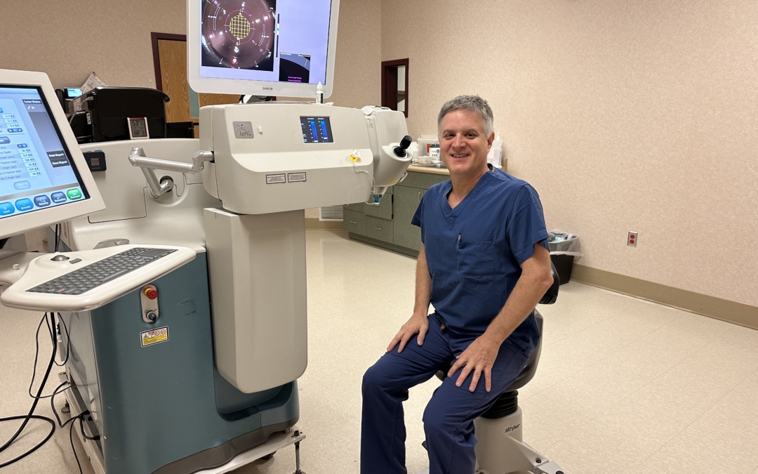
Laser cataract surgery treats cataracts just like traditional surgery — but with advanced laser precision and reduces astigmatism
Instead of a blade, a femtosecond laser performs key steps, including:
- Creating precise corneal incisions
- Opening the lens capsule
- Softening the cataract
- Making precise arcuate incisions to reduce astigmatism
Key Advantage:
Laser cataract surgery can help correct astigmatism during the procedure, something traditional blade-based surgery does not.
The Bottom Line
- LASIK reshapes the cornea to correct refractive errors.
- Traditional cataract surgery removes a cloudy lens using manual surgical techniques.
- Laser cataract surgery removes the cloudy lens with laser precision and can correct astigmatism at the same time.
The right procedure depends on your age, eye health, and visual goals. A comprehensive eye evaluation is the best way to determine which option is right for you
https://www.erielasereye.com/our-services-lasik-eye-surgery/lasik-prk-eye-surgery/
https://www.erielasereye.com/our-services-lasik-eye-surgery/cataract-surgery-with-dr-haverly/
https://www.aao.org/eye-health/treatments/lasik
by Robert Haverly | Jan 18, 2026 | News
Glaucoma: The Silent Thief of Sight
Glaucoma is a group of eye diseases that damage the optic nerve, which carries visual information from the eye to the brain. It is one of the leading causes of irreversible vision loss worldwide. Because glaucoma often has no early symptoms, regular eye exams are essential for early detection and vision preservation.
What Is Glaucoma?
The eye constantly produces a clear fluid called aqueous humor. When this fluid does not drain properly, intraocular pressure (IOP) may rise, leading to optic nerve damage over time. Vision loss from glaucoma is typically gradual and painless, which is why it is often called the “silent thief of sight.”
Types of Glaucoma
Open-angle glaucoma is the most common form and develops slowly as the eye’s drainage system becomes less efficient.
Angle-closure glaucoma is less common but more urgent, occurring when the drainage angle becomes blocked, potentially causing eye pain, blurred vision, halos around lights, nausea, or redness.
Other forms include normal-tension and secondary glaucoma.
Who Is at Risk?
Risk factors for glaucoma include age over 40, family history, African American, Hispanic or Asian ancestry, diabetes, high blood pressure, prior eye injury, and long-term steroid use.
Why Early Detection Matters
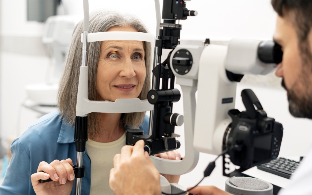
Early glaucoma usually has no warning signs. Comprehensive eye exams allow your ophthalmologist to measure eye pressure, evaluate the optic nerve, test peripheral vision, and detect damage before vision loss occurs. Vision lost from glaucoma cannot be restored, but progression can often be prevented.
Treatment Options
Treatment focuses on lowering eye pressure to protect the optic nerve.
Medications
Prescription eye drops are often the first step to reduce eye pressure.
Laser Treatment
Argon Laser Trabeculoplasty (ALT) and Selective Laser Trabeculoplasty (SLT) improve fluid drainage and may reduce or eliminate the need for drops in some patients.
Surgery
When medications and laser treatment are not sufficient, surgery may be recommended.
Minimally Invasive Glaucoma Surgery (MIGS) offers a safer, less invasive option for patients with mild to moderate glaucoma, often combined with cataract surgery.
Trabeculectomy is a traditional surgery used for moderate to advanced glaucoma to create a new drainage pathway.
Seton or tube shunt implants are used in complex or advanced cases to help control eye pressure.
Living With Glaucoma
Glaucoma is usually a lifelong condition requiring ongoing monitoring and treatment. With proper care and follow-up, most patients can preserve vision and maintain an active lifestyle.
https://www.erielasereye.com/additional-service/glaucoma
https://www.aao.org/eye-health/diseases/what-is-glaucoma
by Robert Haverly | Jan 1, 2026 | News
Macular Degeneration Treatment
Age-related macular degeneration (AMD) is a leading cause of central vision loss in adults over 50. With early diagnosis, advanced testing, and appropriate treatment, many patients are able to preserve useful vision.
At Laser Eye Surgery of Erie, Dr. Robert Haverly provides comprehensive evaluation, advanced diagnostic imaging, and personalized treatment for dry and wet macular degeneration.
What Is Macular Degeneration?
Macular degeneration affects the macula, the central part of the retina responsible for sharp, detailed vision. Common symptoms include:
- Blurry or distorted central vision
- Dark or missing spots in the center of vision
- Difficulty reading or recognizing faces
- Peripheral vision typically remains intact.
Testing and Diagnosis of Macular Degeneration
Accurate diagnosis and ongoing monitoring of AMD rely on advanced retinal imaging technologies, including:
Optical Coherence Tomography (OCT)
OCT is a non-invasive imaging test that provides detailed cross-sectional views of the retina. It helps detect:
- Retinal fluid or swelling
- Thinning or damage to retinal layers
- Early signs of wet AMD or geographic atrophy
OCT is commonly used at follow-up visits to monitor disease progression and response to treatment.
OCT Angiography (OCT-A)
OCT-A is an advanced, non-invasive imaging technique that visualizes blood flow in the retinal and choroidal vessels without the need for dye. It is especially useful for:
- Detecting early or subtle abnormal blood vessels
- Identifying wet AMD before fluid appears
- Monitoring response to injections for AMD
Fluorescein Angiography (FA)
Fluorescein angiography uses a special dye and camera system to evaluate retinal blood flow. It is particularly helpful for:
- Confirming wet AMD
- identifying leaking or abnormal blood vessels
- Guiding treatment decisions in complex cases
Fundus Autofluorescence (FAF)
Fundus autofluorescence imaging highlights metabolic changes in retinal cells and is especially valuable for:
- Evaluating geographic atrophy (advanced dry AMD)
- Tracking progression of retinal cell loss
- Identifying areas at risk before vision changes occur
Dry Macular Degeneration
Dry AMD is the most common form and usually progresses slowly.
Early & Intermediate Dry AMD
Most patients with early or intermediate dry AMD do not require injections. Management typically includes:
- AREDS2 eye vitamins (when appropriate)
- UV-protective sunglasses
- Routine monitoring with retinal imaging
- Healthy lifestyle habits, including not smoking
- Many patients maintain stable vision for years with these measures alone.
Geographic Atrophy (Advanced Dry AMD)
Geographic atrophy (GA) is an advanced form of dry AMD that causes progressive central vision loss.
Injection treatments for GA include:
Syfovre® (pegcetacoplan) – FDA approved
Izervay® (avacincaptad pegol) – FDA approved
These medications can slow disease progression and help preserve remaining vision. Injections are typically given every one to two months, depending on the treatment plan. They do not restore lost vision.
Wet Macular Degeneration
Wet AMD is less common but more aggressive. It develops when abnormal blood vessels grow beneath the retina and leak fluid or blood.
Injection Treatments for Wet AMD
Wet AMD is treated with anti-VEGF injections, including:
- Lucentis® (ranibizumab) – FDA approved
- Eylea® (aflibercept) – FDA approved
- Vabysmo® (faricimab) – FDA approved
- Avastin® (bevacizumab) – commonly used off-label
Some medications also have biosimilar versions, which may be used based on medical needs and insurance coverage. Most wet AMD injections are administered every one to two months, especially early in treatment, with schedules adjusted based on response. With timely treatment, many patients experience stabilized vision, and some see improvement.
Ongoing Monitoring Is Essential
Macular degeneration requires regular follow-up using advanced imaging to detect changes early and guide long-term treatment decisions.
Trusted Macular Degeneration Care in Erie
With over 30 years of ophthalmology experience, Dr. Robert Haverly provides trusted, personalized care for macular degeneration at Laser Eye Surgery of Erie.
https://www.erielasereye.com/additional-service/macular-degeneration/
https://www.aao.org/eye-health/diseases/amd-macular-degeneration
