PTK and Amniotic Tissue Graft
Home /
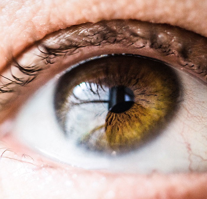
Phototherapeutic Keratectomy (PTK)
Phototherapeutic Keratectomy (PTK) and Amniotic Membranes with bandage contacts play an important role in treating corneal dystrophies and keratitis.
Corneal dystrophies, such as Epithelial Basement Membrane Dystrophy (EBMD), and keratitis are serious conditions that can cause vision impairment and discomfort. However, advancements in ophthalmology have introduced innovative treatments to address these conditions effectively. This essay explores two treatment modalities: Phototherapeutic Keratectomy (PTK) for corneal dystrophies like EBMD and the utilization of amniotic membranes with bandage contacts in managing keratitis. Both approaches have shown significant efficacy in improving visual outcomes and promoting corneal healing.
Phototherapeutic Keratectomy (PTK) for Corneal Dystrophies
PTK is a minimally invasive surgical technique performed using an excimer laser. It involves removing the superficial layer of the cornea, known as the epithelium, and reshaping the underlying corneal tissue. This procedure eliminates irregularities and abnormal deposits, providing benefits for corneal dystrophies like EBMD.
PTK offers several benefits in treating EBMD. Firstly, it resolves recurrent corneal erosions by removing the irregular epithelium responsible for these erosions. This promotes better adhesion between the epithelium and underlying corneal tissue, reducing the risk of corneal erosions and associated pain and discomfort. Secondly, PTK smooths the corneal surface by eliminating irregularities and abnormal deposits in the epithelial basement membrane. This leads to a reduction in visual disturbances such as glare and blurry vision, resulting in improved visual acuity and clarity for patients.
Preoperative considerations for PTK involve comprehensive patient evaluation, including corneal topography and assessment of visual acuity. These evaluations determine the extent and severity of EBMD and assist in planning the surgical approach. Patient education is crucial to establish realistic expectations regarding visual outcomes and postoperative care.
Postoperative management include eye drops to prevent infection and reduce inflammation. Lubricating eye drops are also recommended to alleviate dryness and promote healing. Regular postoperative visits monitor corneal healing, visual acuity, and symptom improvement, with necessary adjustments to medications or further interventions based on patient progress.
Home /
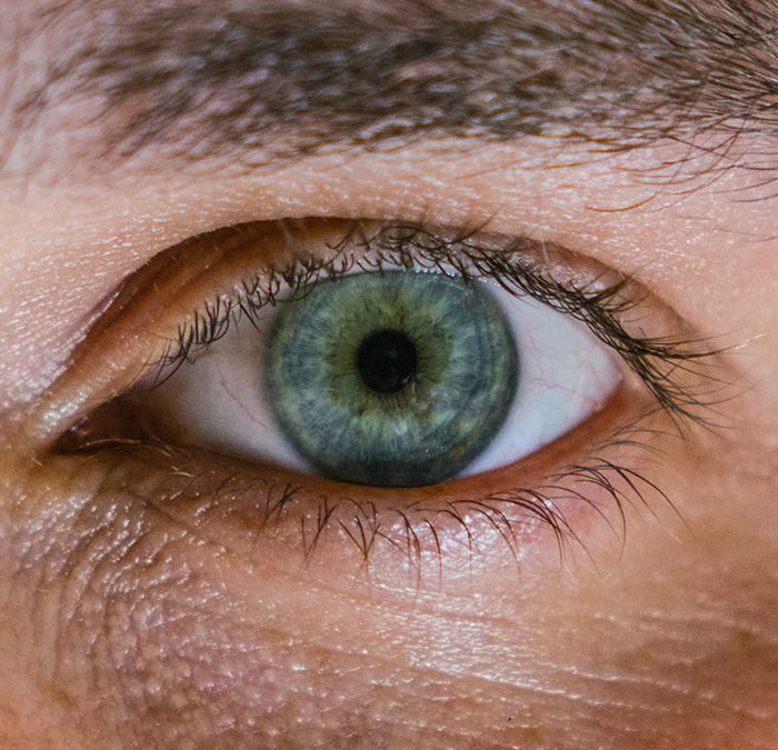
Amniotic Tissue Graft
Amniotic Membrane Graft
Common causes of keratitis include corneal dystrophies, such as epithelial basement membrane disease (EBMD), dry eyes, exposure keratopathy, allergies, and contact lenses. When keratitis develops prompt and appropriate treatment is needed to prevent vision loss. Amniotic membranes are derived from the placenta have gained recognition as a valuable treatment modality for keratitis. When combined with bandage contacts, they provide a protective environment for the cornea, promoting healing and improving visual outcomes.
The treatment process begins with the application of amniotic membrane directly onto the affected cornea. The membrane adheres to the corneal surface, acting as a barrier to protect the cornea and stimulate healing. A bandage contact lens, typically made of soft silicone hydrogel material, is then placed over the amniotic membrane. This lens provides additional protection, reduces friction, and enhances patient comfort. The contact lens stays in your eye until it is removed by us. We usually keep the contact lens in place for 3 to 7 days. However, if it comes out before this time do not place it back into your eye.
Amniotic membranes with bandage contacts offer several benefits in the treatment of keratitis. They promote healing by releasing growth factors and cytokines that stimulate tissue repair, reduce inflammation, and inhibit scarring. Additionally, they alleviate pain and discomfort associated with keratitis by acting as a protective barrier and reducing mechanical irritation. The antimicrobial properties of amniotic membranes prevent and control infections, while their anti-inflammatory and anti-fibrotic properties minimize scarring and complications.
Conclusion
PTK and the utilization of amniotic membranes with bandage contacts represent significant advancements in the treatment. This has become a common, non-painful, way to treat EBMD with keratitis.

Laser Eye Surgery of Erie
Additional Services
PTK and Amniotic Tissue Graft
Home Phototherapeutic Keratectomy (PTK) Phototherapeutic Keratectomy (PTK) and Amniotic Membranes with bandage contacts play an important role in treating…
read more
Corneal Transplants
Corneal transplants are required for a number of corneal diseases and a variety of other reasons. Before and after transplant,…
read more
Dry Eye
Dry eye occurs when people don’t produce enough tears or the right quality of tears to keep their eyes healthy…
read more
Diabetic Services
Diabetic retinopathy is a disease that affects diabetic people and eventually leads to blurry, distorted vision and blindness. When people…
read more
Macular Degeneration
Age-related macular degeneration (AMD) is a deterioration or breakdown of the eye’s macula. The macula is a small area in…
read more
Glaucoma Surgery and MIGS
iStent works like the stents used to prevent heart attacks and strokes. When blood vessels get clogged, a stent creates…
read more
Retinal Surgery
The retina is a thin sheet of nerve tissue in the back of the eye where light rays are focused…
read more
Keratoconus & Corneal Cross Linking
Keratoconus, often referred to as ‘KCN’, is a non-inflammatory eye condition in which the typically round dome-shaped cornea progressively thins…
read more
Glaucoma
Although glaucoma is the leading cause of blindness in Americans, early detection can prevent you from losing your eyesight. In…